Imaged parent items


New Approaches to the Measurement of MS Brain Pathophysiology
Finding a cure for MS requires better definition of the relationship between the pathophysiology of the central nervous system (CNS) and the clinical course. A routine MRI is very helpful for diagnosis but less so for prognosis. Presently there is no single imaging modality that can sufficiently link future neurologic function to CNS pathophysiology. Further, it is unlikely that using only one MS neuroimaging modality
can lead to a full picture of the structure/function relations of MS on the brain and spinal cord.
Fortunately, a multimodal simultaneous neuroimaging approach is now possible at Stony Brook. Thanks to a gift from Robert and Lisa Lourie, Stony Brook Medicine recently obtained a simultaneous Positron Emission Tomography (PET) /MRI scanner, the 3 Tesla Siemens Biograph mMR, one of very few in use worldwide. Since PET and MRI images are acquired simultaneously, in performing research studies we can examine the brain using four imaging modalities simultaneously, without additional patient burden. These imaging modalities include (a) structural MRI (sMRI) of grey and white matter cerebral volume, (b) diffusion spectrum imaging (DSI) of white matter (structural connectivity) disruptions, (c) functional MRI (fMRI) of variability in the functional connectivity of neural pathways, and (d) PET to measure brain metabolism. Application of these modalities provide an opportunity to link structure and function.
At Stony Brook we are about to launch a study with the new MRI/PET scanner that will examine, during one single scanning procedure, all four of these methods in a single person. The goal of this research is to link intensive analysis of cognitive performance and its treatment with structure and neuronal function. Neuroimaging measures and cognitive performance will also be combined with measures of day to day functioning, fatigue, and other aspects of neurological function. We will monitor individuals longitudinally so that we can address the important question of how even subtle effects of MS on brain structure/function affect overall neurologic and cognitive wellbeing and we will ask the question whether how targeted interventions can partially reverse the effects of MS.
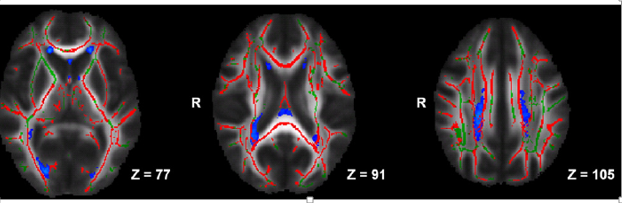
Recently we published a study comparing just one neuroimaging modality (Diffusion Tensor Imaging (DTI) with cognitive measures on individuals with MS and those without the disease. We were able to create maps of lesion probability (blue), changes in water diffusion along the whiter matter tracts (red), and link these to performance on cognitive tests.
Disruption of white matter tracts and presence of lesions in MS
Using multiple imaging modalities, we hope to learn the mechanisms of action of various interventions designed to improve MS symptoms. At the moment we are in the also in the process of combining a treatment study of cognition with new neuroimaging approaches. Future studies will focus on other treatment strategies.
We are optimistic that these technological advances will provide answers to how MS develops and, ultimately, how it can be stopped.

Stony Brook University Hospital
101 Nicolls Road Stony Brook, NY 11794
(631) 444-4000